Acute inflammation of the liver due to heavy alcohol use, usually after prolonged drinking, characterized by jaundice and liver enzyme elevation (AST > ALT, typically AST:ALT ≈ 2:1).
Severe alcoholic hepatitis has a high mortality (≈30–50% 28-day mortality if untreated). It's a common reason for hospitalization in heavy drinkers and a classic exam scenario (e.g., recognizing AST:ALT ratio). Prompt diagnosis and treatment (e.g., steroids) can be life-saving, and preventing progression to cirrhosis requires early intervention and alcohol abstinence.
Middle-aged heavy drinker (often decades of alcohol use, e.g., >8 drinks/day) with acute illness after a binge or continued drinking. Frequently malnourished and may have other signs of alcohol abuse (e.g., parotid enlargement, Dupuytren contractures).
Presents with jaundice (often developing over weeks), fever, fatigue, anorexia, and right upper quadrant pain. Exam typically shows hepatomegaly (liver enlarged and tender). May also see ascites or encephalopathy if severe.
Labs: moderate AST and ALT elevation (often 100–300 U/L range, AST about 2× ALT). Bilirubin is elevated (often >5 mg/dL) and INR prolonged (coagulopathy) in severe cases. Leukocytosis (↑WBC) is common, and GGT is often high (reflecting alcohol use).
Exclude other causes: test for viral hepatitis (HAV, HBV, HCV serologies), check for drug-induced liver injury (medication history, acetaminophen level if overdose possible), and obtain imaging (e.g., ultrasound) to rule out biliary obstruction or other pathology. Liver biopsy is diagnostic (showing neutrophils and Mallory bodies), but usually only done if the diagnosis is uncertain.
Identify heavy alcohol use in history (usually years of excessive drinking). Assume honesty is crucial when discussing alcohol intake.
Check LFTs: look for AST:ALT ~2:1 (AST typically <300 U/L, ALT <200), elevated bilirubin, and high GGT (supports alcohol use). Also check CBC (often ↑WBC), INR/PT (prolonged if severe), and albumin (low in chronic liver disease/malnutrition).
Rule out alternative causes of hepatitis: order viral hepatitis panel (A, B, C), consider tests for autoimmune hepatitis (ANA, SMA), Wilson disease (ceruloplasmin) or hemochromatosis (iron studies) if indicated. Review medications and toxins for possible DILI.
Perform abdominal ultrasound (or CT) to assess liver size (often enlarged with fatty change) and exclude biliary obstruction or masses. If ascites is present, tap it to check for infection (SBP).
Assess disease severity with prognostic scores: calculate Maddrey Discriminant Function (DF) = 4.6 × (patient's PT - control PT) + total bilirubin (mg/dL). DF ≥ 32 indicates severe hepatitis with ~30–50% short-term mortality. Also calculate MELD score (uses bilirubin, INR, creatinine; MELD >21 is another marker of severe disease).
If considering steroid therapy, first screen for infection (blood/urine cultures, chest X-ray, etc.) since active infection contraindicates steroids. Start treatment promptly if criteria met.
Condition
Distinguishing Feature
Acute viral hepatitis
very high ALT (often >1000), history of viral exposure (travel, IV drugs, etc.), no significant alcohol use
Drug-induced liver injury (DILI)
history of toxin/medication exposure (e.g., acetaminophen overdose or other hepatotoxic drugs); pattern varies but ALT often very high
Non-alcoholic fatty liver disease (NAFLD/NASH)
similar fatty liver pathology but occurs in non-drinkers with metabolic syndrome; usually chronic mild ALT elevation rather than acute hepatitis
Absolute alcohol abstinence is critical – the only way to allow liver healing. Provide counseling and support (rehab, support groups); relapse prevention is key.
Nutritional support: high-protein, high-calorie diet and vitamins (e.g., folate, thiamine). Many patients are malnourished; replete deficiencies and monitor electrolytes (refeeding syndrome risk).
If severe AH (e.g., MDF ≥32 or MELD >21) with no contraindications, start corticosteroids (typically prednisolone 40 mg daily for 4 weeks). This improves short-term survival. Monitor response via Lille score at 7 days: if Lille <0.45, continue steroids; if ≥0.45 (poor responder), stop steroids (no benefit).
If steroids are contraindicated (e.g., active infection, GI bleeding) or not tolerated, consider pentoxifylline 400 mg TID as an alternative (mixed evidence, less effective than steroids).
Manage complications: use lactulose for encephalopathy, antibiotics for infections, beta-blockers/endoscopy for varices if present, and diuretics/paracentesis for ascites as needed. For patients who fail medical therapy and remain very ill, early liver transplant can be considered in select cases (first episode, committed to sobriety).
AST = A Scotch and Two-beer (2:1 AST:ALT) is a mnemonic for alcohol-related liver injury (AST about 2× ALT). In contrast, viral hepatitis typically causes ALT > AST.
Transaminase levels in alcoholic hepatitis are usually moderate (AST/ALT rarely >500 U/L) due to alcohol's effect on enzyme production (vitamin B6 deficiency). Much higher levels (thousands) suggest other causes (viral or acetaminophen injury).
Mallory-Denk bodies (intracytoplasmic keratin inclusions) on liver biopsy are classically associated with alcoholic hepatitis (though not specific, also seen in NASH).
Always check for infections in severe alcoholic hepatitis (SBP, pneumonia, UTI) – these patients are prone to sepsis, and untreated infection plus steroids can be fatal.
New fever or hypotension in an AH patient → suspect sepsis (common in severe AH); evaluate immediately (cultures, antibiotics) before or during steroid therapy.
GI bleeding (e.g., hematemesis or melena) in alcoholic hepatitis indicates variceal hemorrhage or peptic ulcer; requires urgent endoscopy and stabilization (this also contraindicates steroid use until bleeding is controlled).
Worsening mental status (hepatic encephalopathy) or asterixis → signifies severe liver failure. These patients need ICU-level care; consider this an indication for transplant evaluation if not improving.
Heavy alcohol use + acute jaundice and AST:ALT ~2:1 → suspect alcoholic hepatitis.
Evaluate with labs (AST, ALT, bilirubin, INR, CBC) and imaging (ultrasound); rule out other causes (viral hepatitis panel, drug screen).
If diagnosis is likely, calculate severity scores (Maddrey DF, MELD). If DF ≥32 or MELD >21, plan to initiate steroids (after checking for infections).
Start prednisolone 40 mg/day for 28 days if severe AH and no contraindications. Re-check patient at 7 days (use Lille score to decide whether to continue steroids).
Ensure strict alcohol abstinence and provide nutritional therapy. Monitor for complications (infections, GI bleeding, encephalopathy) and treat/support accordingly. Arrange follow-up for addiction treatment and consider transplant for non-responders.
A 50‑year‑old heavy drinker with 2 weeks of jaundice, fever, and tender hepatomegaly; labs show AST ~200, ALT ~90 (AST:ALT ≈2:1), elevated bilirubin and INR → acute alcoholic hepatitis (recognize alcohol pattern and treat severe cases with steroids).
Patient with acute alcoholic hepatitis has Maddrey DF 40 and new-onset encephalopathy → severe AH with poor prognosis; the next step is to start corticosteroids (prednisolone) after ruling out infection.
Histology vignette: Liver biopsy showing neutrophilic inflammation and Mallory bodies in a patient with long-term alcohol use → alcoholic steatohepatitis (alcoholic hepatitis).
Case 1
A 45‑year‑old man with a 20-year history of heavy alcohol use (8–10 beers per day) presents with 2 weeks of jaundice and abdominal pain.
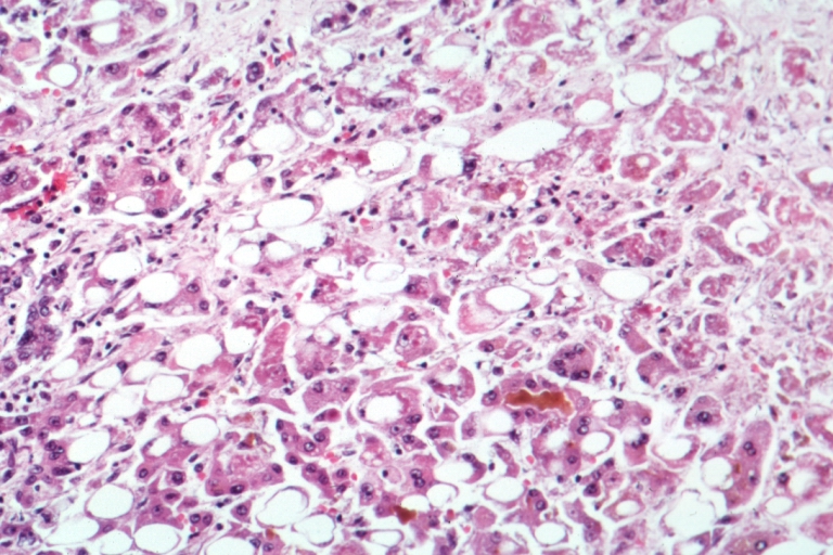
Microscopic liver tissue (H&E stain) showing features of alcoholic hepatitis: fat droplets, hepatocyte ballooning/necrosis, and a characteristic **Mallory body** (pink inclusion).