Holoprosencephaly
High-YieldFree study guide for medical students and educators
Holoprosencephaly (HPE) is a congenital forebrain malformation where the embryonic prosencephalon fails to divide into two separate hemispheres. The result is a single midline cerebral structure (monoventricle) often with craniofacial midline defects.
- HPE links early embryology to classic exam clues. Severe facial defects (cyclopia, proboscis) imply alobar HPE, while subtle signs (single maxillary incisor) hint at a microform. It is often associated with trisomy 13 (Patau syndrome) and SHH pathway mutations, so recognition prompts genetic evaluation.
- Prenatal US: single large brain ventricle and fused hemispheres, absent interhemispheric fissure.
- Infant: microcephaly, seizures, and midline facial anomalies (cyclopia/synophthalmia, proboscis, cleft lip/palate).
- Child (mild): isolated facial findings (single central incisor, hypotelorism) with learning delays.
- MRI: fused cerebral hemispheres with absent corpus callosum; a single ventricle is seen.
- Genetics: karyotype/microarray often shows trisomy 13; test for SHH, ZIC2, SIX3 mutations.
- If HPE is suspected on fetal US, confirm with fetal MRI to define severity.
- Obtain karyotype/microarray (evaluate for trisomy 13/18) and consider gene panels (SHH, SIX3, etc.).
- Postnatal MRI to classify (alobar, semilobar, lobar, MIH).
- Evaluate for complications: assess pituitary function (diabetes insipidus, adrenal insufficiency), manage seizures, consult neurosurgery for hydrocephalus if present.
| Condition | Distinguishing Feature |
|---|---|
| Agenesis of corpus callosum | Two separate hemispheres (no fused ventricle) but missing corpus callosum; normal appearance of lateral ventricles. |
| Septo-optic dysplasia | Optic nerve hypoplasia and absent septum pellucidum; milder midline defect without hemispheric fusion. |
| Hydranencephaly | Destructive loss of cerebral hemispheres (CSF-filled sac); macrocephaly and no brain tissue, differing from developmental fusion. |
- HOLO prosencephaly = whole forebrain stays as one (one ventricle).
- Sonic Hedgehog (SHH) mutations → HPE (Hedgehog → HPE mnemonic).
- Cyclops (one eye) = think alobar HPE (one-ventricle brain).
- 13 (Patau) has HPE; recall '1 eye, 3 halves' for cyclopia.
- Suspect HPE if facial midline defect or abnormal fetal brain ultrasound.
- Perform fetal MRI to evaluate brain structure.
- Obtain genetic studies (karyotype/microarray for trisomies; HPE gene panel for SHH, SIX3, etc.).
- Manage complications: neurosurgical consult for hydrocephalus, endocrine for pituitary issues, and genetic counseling.
- Prenatal US: fused thalami + single ventricle → Holoprosencephaly.
- Newborn with cyclopia (one eye) and proboscis → Alobar HPE.
- Infant with cleft lip and hypotelorism, MRI shows partial hemispheric fusion → Semilobar HPE.
- Toddler with a single central incisor and developmental delay → HPE microform.
A 22-week fetal ultrasound shows a single large midline ventricle with fused thalami. The fetal face has a small proboscis above where the eyes should be.
A 2-year-old has developmental delay and facial anomalies: a single central incisor and cleft palate. Brain MRI is essentially normal with no midline structures missing.
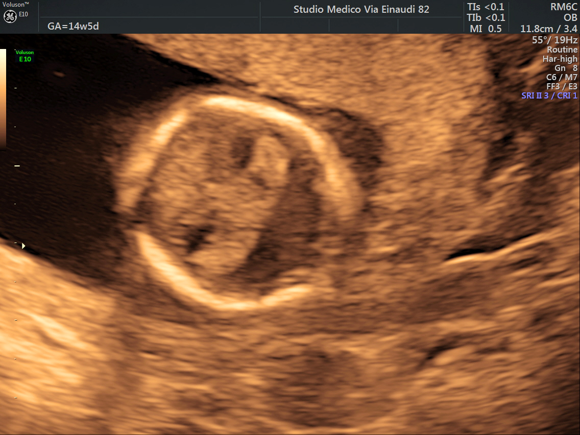
Ultrasound of 14-week fetus with holoprosencephaly: a single midline ventricle (monoventricle) and absent interhemispheric fissure
image credit