Aggressive malignancy of the mesothelial lining of serous cavities (pleura >> peritoneum), usually due to asbestos exposure; pleural mesothelioma characteristically encases the lung in a thick rind.
Lethal cancer (median survival ~1 year) with a long latency period. Exemplifies the severe long-term risk of asbestos exposure and often appears on exams as the classic asbestos-associated malignancy (distinct from smoking-related lung carcinoma).
Pleural mesothelioma: older adult with chronic asbestos exposure history (e.g., shipyard, construction) presenting with insidious dyspnea, chest wall pain, cough, and weight loss. Usually a unilateral pleural effusion (often hemorrhagic) is present; imaging shows diffuse pleural thickening and a mass encasing the lung.
Peritoneal mesothelioma: progressive abdominal distension, pain, and ascites due to diffuse peritoneal tumor spread. Can occur after heavy asbestos exposure (ingested fibers). Rarely, mesothelioma arises in the pericardium (causing effusive-constrictive pericarditis) or in the tunica vaginalis of the testes (presents as a hydrocele).
Unilateral pleural disease in an asbestos-exposed patient should raise suspicion for mesothelioma even decades after exposure – pursue evaluation rather than attributing to benign causes.
Obtain a thoracoscopic pleural biopsy for definitive diagnosis (pleural fluid cytology is often non-diagnostic, with <30% sensitivity).
Immunohistochemistry (IHC) helps distinguish mesothelioma from adenocarcinoma: mesothelioma cells typically stain positive for calretinin, WT-1, and cytokeratin 5/6, and are negative for adenocarcinoma markers like CEA or TTF-1.
For peritoneal presentations, exclude more common causes of peritoneal carcinomatosis (e.g., ovarian or colon cancer metastases) by appropriate imaging and biopsy.
Condition
Distinguishing Feature
Metastatic lung adenocarcinoma
more common lung cancer (especially in smokers) that can involve pleura; glandular on histology, often CEA/TTF-1 positive, usually presents with a discrete lung mass
Peritoneal carcinomatosis (ovarian/GI cancer)
malignancy spread from another primary (e.g., ovarian carcinoma) causing diffuse peritoneal implants; usually no asbestos history and different tumor markers
Benign asbestos pleural disease
pleural plaques or benign asbestos effusions without invasive tumor; history of asbestos exposure but no malignancy (no pleural thickening rind or mass)
Early-stage (localized) disease: aggressive surgery (pleurectomy/decortication or extrapleural pneumonectomy) combined with chemotherapy (cisplatin + pemetrexed) ± radiation (trimodal therapy) can be attempted in select patients.
Advanced or unresectable disease: systemic therapy is mainstay – platinum + pemetrexed chemotherapy (often with bevacizumab) or immunotherapy (combined checkpoint inhibitors like nivolumab + ipilimumab). Palliative pleurodesis (or indwelling pleural catheter) helps control recurrent effusions.
Peritoneal mesothelioma: optimal management is cytoreductive surgery with HIPEC (heated intraperitoneal chemotherapy), which can significantly prolong survival in eligible patients.
Exam tip: asbestos exposure + pleural tumor (especially with effusion) → think mesothelioma (unlike lung cancer, mesothelioma risk is not linked to smoking).
Pathology buzzword: mesothelioma cells have long, slender microvilli on electron microscopy (vs adenocarcinoma cells with shorter microvilli).
Pleural mesothelioma tends to form a diffuse tumor rind encasing the lung rather than a single lung nodule.
In an asbestos-exposed patient, a persistent unilateral pleural effusion or unexplained pleural thickening is a red flag for mesothelioma – prompt imaging and biopsy are essential (don't assume it's benign).
Mesothelioma involving the pericardium can lead to cardiac tamponade – watch for hypotension, JVD, distant heart sounds; if present, urgent pericardial drainage is required.
Imaging: get chest X-ray (may show pleural plaques or effusion) then CT chest to evaluate pleural thickening, masses, and extent of disease.
Thoracentesis (diagnostic & therapeutic) if effusion is present – fluid is typically exudative and often hemorrhagic, but cytology is usually non-diagnostic.
Thoracoscopy with pleural biopsy for definitive diagnosis (required to confirm mesothelioma histologically and via IHC, distinguishing it from adenocarcinoma).
Stage the disease (CT/PET ± MRI); involve a multidisciplinary team to determine treatment – surgery for early-stage fit patients vs chemotherapy/immunotherapy for advanced disease; integrate palliative care early for symptom management.
Older man with a history of shipyard asbestos exposure presents with chest pain, dyspnea, and unilateral pleural effusion; imaging shows pleural thickening encasing one lung → malignant pleural mesothelioma.
Patient with a history of asbestos exposure develops abdominal distension and ascites; exploratory laparoscopy reveals diffuse peritoneal tumor implants → malignant peritoneal mesothelioma.
Case 1
A 72-year-old man with a 30-year history of shipyard work (and known asbestos exposure) presents with 6 months of progressive shortness of breath and chest pain.
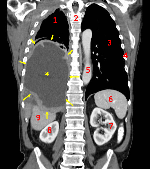
Coronal CT scan of the chest showing malignant pleural mesothelioma (tumor encasing the left lung, with a pleural effusion).